What it means
Understanding lumbar spondylolisthesis
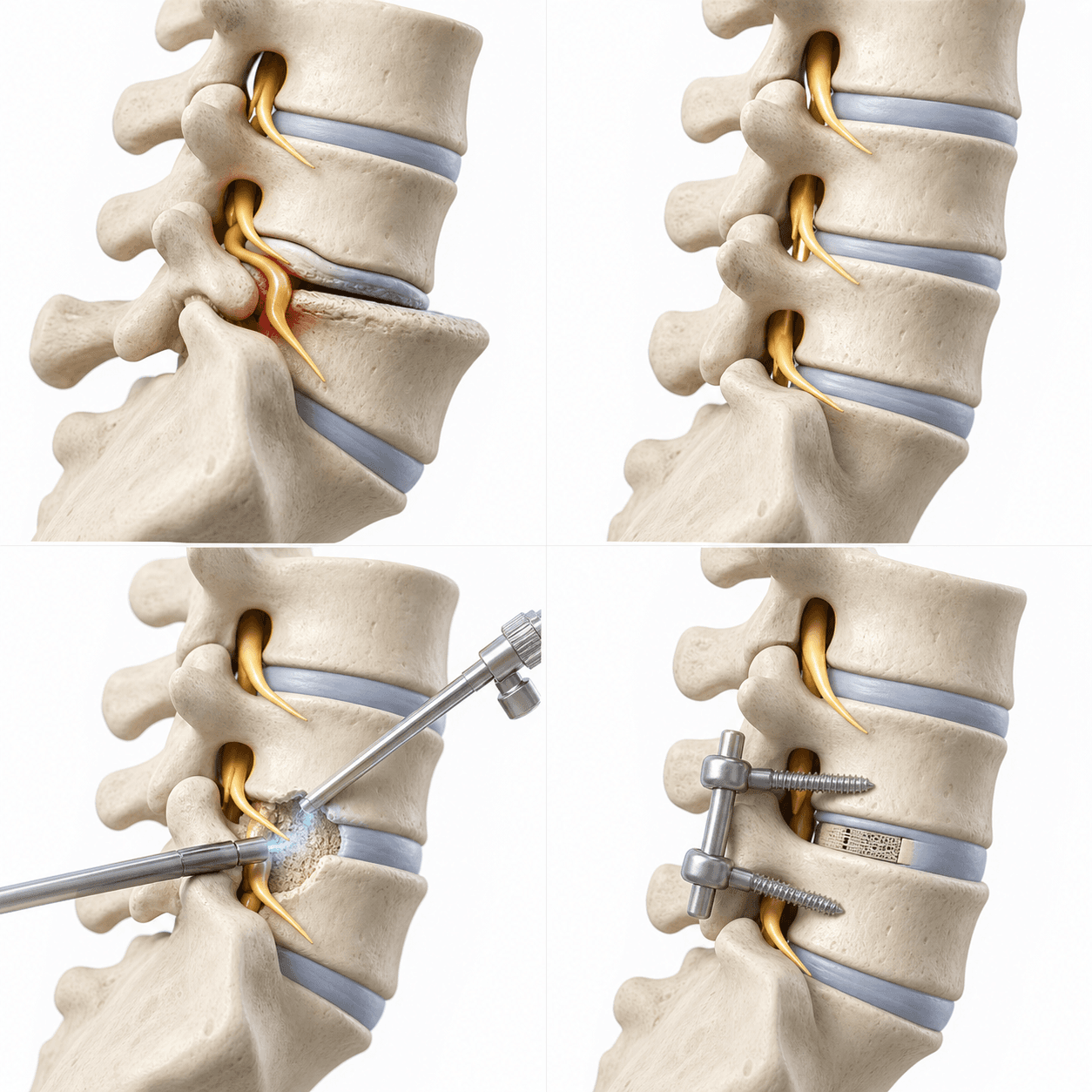
Degenerative lumbar spondylolisthesis occurs when one vertebra slips forward relative to the vertebra below as the joints and discs change over time. Symptoms may result from associated nerve compression or clinically relevant instability rather than from the amount of slippage alone.
Common patterns
Symptoms to discuss
- Low-back pain with standing, lifting, or position changes
- Leg pain, numbness, heaviness, or weakness from associated nerve compression
- Reduced walking or standing tolerance
- Symptoms that vary with posture, activity, and the degree of instability
Clinical correlation
How the condition is evaluated
- Assessment of back symptoms, leg symptoms, walking, and daily function
- Neurologic examination of strength, sensation, and reflexes
- Standing X-rays to assess alignment and the degree of slippage
- Flexion-extension X-rays when appropriate to assess motion, plus MRI to identify nerve compression
First-line options
Non-surgical care
- Medication and activity modification
- Guided trunk and lower-limb exercise
- Selected injection treatment when appropriate
- Monitoring for changes in walking tolerance, leg strength, or instability-related symptoms
Individual decision
When surgery may be discussed
- Surgery may be considered when leg symptoms, weakness, or walking limitations persist despite non-surgical care.
- Imaging should demonstrate nerve compression or instability that is clinically relevant to the symptoms.
- Decompression alone may be considered when stability can be maintained.
- If clinically relevant instability is present, UBE-assisted transforaminal lumbar interbody fusion (UBE-TLIF) may be considered. Fusion is not required for every patient with spondylolisthesis.
Procedure concept
Decompression Alone or Decompression with Fusion
The operative plan begins with the reason for surgery, not the access method. Biportal endoscopic decompression may be considered when decompression alone is appropriate. When instability, foraminal collapse, or the planned decompression requires stabilization, UBE-TLIF or another fusion approach may be discussed.
- 1The symptomatic nerve target and degree of instability are reviewed.
- 2The surgeon determines whether decompression can be performed without destabilizing the segment.
- 3If decompression alone is selected, the planned compressive tissue is removed.
- 4If fusion is required, disc-space preparation, an interbody implant, bone graft, and fixation may be added according to the surgical plan.
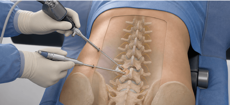
This AI-generated 3D medical illustration is provided for general patient education. It is not an image of an actual patient, an actual operation, or a before-and-after result, and it does not predict an individual treatment outcome. Anatomy, disease patterns, and surgical steps vary from person to person.
General planning ranges
Recovery and return to activity
| Activity | Planning range | What affects timing |
|---|---|---|
| Desk or remote work | Decompression: about 2–4 weeks; fusion: about 6–12 weeks | Begin with shorter periods and increase gradually. |
| Driving or field sales | Decompression: about 3–6 weeks; fusion: about 6–12 weeks | Safe braking, sitting tolerance, and medication status matter. |
| Light standing work | Decompression: about 4–8 weeks; fusion: about 8–12 weeks | Walking endurance and neurologic function guide progression. |
| Heavy or repetitive work | Decompression: often 12 weeks or later; fusion: often 3–6 months | Imaging, strength, job demands, and the fusion plan must be reviewed. |
These are general planning ranges, not fixed deadlines. Return to driving, work, lifting, or overhead activity should be individualized according to the procedure, wound status, pain medication use, neurologic and functional recovery, imaging findings, and actual job demands.
Informed decision-making
Possible risks and limitations
- Infection, bleeding, or risks related to anesthesia
- Dural tear and spinal fluid leakage
- Nerve injury or new neurologic symptoms
- Persistent, recurrent, or incompletely improved symptoms
- The need for another procedure or a different surgical approach
- Instability after decompression
- For fusion: nonunion, implant-related problems, or adjacent-segment stress
Bring to the appointment
Questions to ask
- Are my symptoms caused by nerve compression, instability, or both?
- Can decompression be performed while maintaining stability?
- What specific finding would make fusion appropriate in my case?
- How would decompression alone and fusion change my recovery plan?
Medical review and sources
This page provides general patient education and does not replace an individual diagnosis or treatment plan. Treatment options, the surgical approach, recovery, and return-to-work timing vary according to the level and location of nerve compression, neurologic findings, overall health, imaging findings, medication use, and actual job demands.
Medically reviewed by Hanjin Jang, MD, Neurosurgeon, Founder and Chief Director, New Standard Hospital. Last reviewed: 2026-07-31.