What it means
Understanding cervical disc herniation
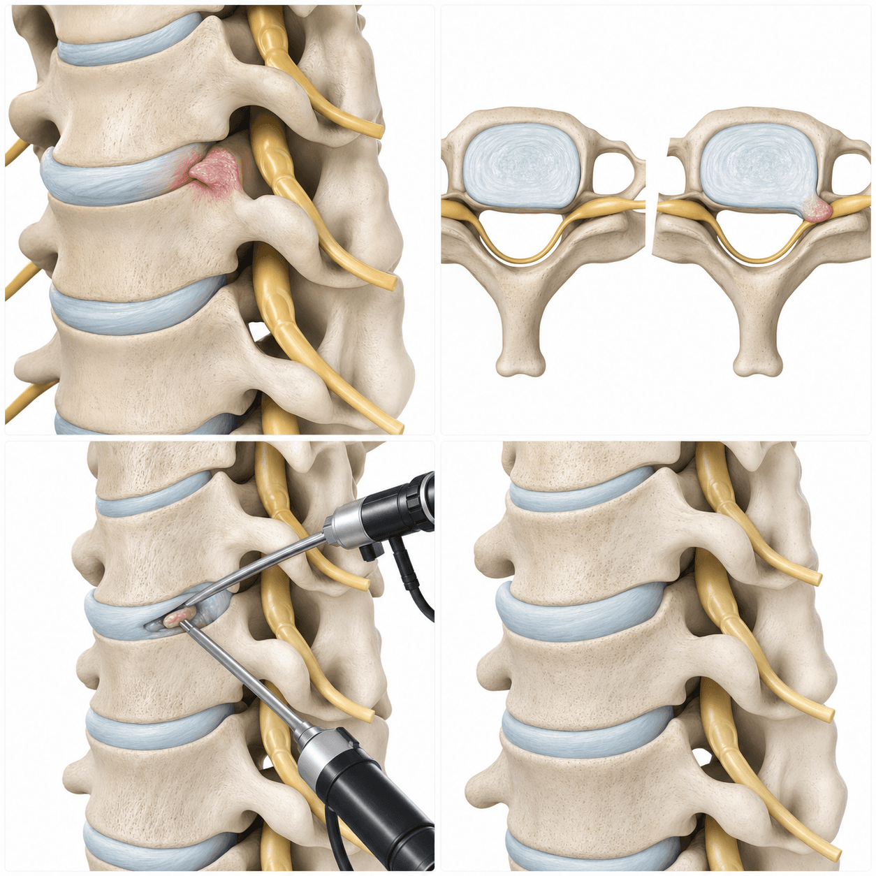
Cervical disc herniation occurs when disc material in the neck irritates or compresses a nerve root and, in some cases, the spinal cord. The location of pain, numbness, or weakness should be consistent with the neurologic examination and imaging findings before a procedure is considered.
Common patterns
Symptoms to discuss
- Neck pain with pain or tingling into the shoulder, arm, or hand
- Numbness or altered sensation in part of the arm or hand
- Weakness in the shoulder, elbow, wrist, or fingers
- Pain that changes with neck position, coughing, or arm movement
Clinical correlation
How the condition is evaluated
- Mapping the distribution of arm pain, numbness, and weakness
- Neurologic examination of strength, sensation, reflexes, hand function, and walking
- MRI to determine whether the side and level of compression match the symptoms
- Review of whether symptoms are improving with medication, guided exercise, activity modification, or selected injections
First-line options
Non-surgical care
- Medication and short-term activity modification
- Guided exercise or physical therapy when appropriate
- Selected injection treatment for symptom control
- Monitoring for new weakness, hand dysfunction, balance problems, or gait change
Individual decision
When surgery may be discussed
- Surgical decompression may be considered when arm pain remains disabling despite appropriate non-surgical care.
- Progressive weakness or signs of spinal cord involvement require prompt evaluation.
- The side and level on imaging should match the symptoms and neurologic findings.
- The surgical approach depends on the location of compression, alignment, stability, and spinal cord involvement; a posterior biportal approach is not appropriate for every cervical disc herniation.
Procedure concept
Posterior Cervical Biportal Endoscopic Foraminotomy and Discectomy
When a posterior biportal approach is appropriate, separate viewing and working portals can be used to enlarge the nerve exit and address selected disc material contributing to nerve-root compression. Other patients may require a different approach.
- 1The side, level, nerve target, and compression pattern are confirmed.
- 2Viewing and working portals are placed through a posterior approach.
- 3The planned portion of the nerve exit is decompressed while the facet joint is preserved as the procedure allows.
- 4Selected disc material may be removed when it is accessible and relevant to the nerve compression.
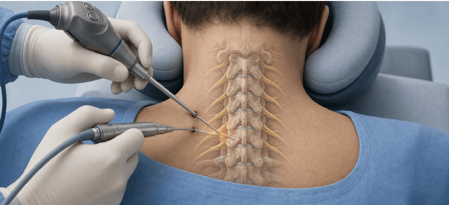
This AI-generated 3D medical illustration is provided for general patient education. It is not an image of an actual patient, an actual operation, or a before-and-after result, and it does not predict an individual treatment outcome. Anatomy, disease patterns, and surgical steps vary from person to person.
General planning ranges
Recovery and return to activity
| Activity | Planning range | What affects timing |
|---|---|---|
| Desk or remote work | About 2–4 weeks | Use an eye-level screen and begin with shorter periods. |
| Driving or field sales | About 3–6 weeks | Neck rotation, field of view, emergency braking, and medication status must be safe. |
| Light standing work | About 4–8 weeks | Begin with tasks that keep the arms closer to the body. |
| Heavy or overhead work | Often 8–12 weeks or later | Strength and neurologic recovery should be reviewed before progression. |
These are general planning ranges, not fixed deadlines. Return to driving, work, lifting, or overhead activity should be individualized according to the procedure, wound status, pain medication use, neurologic and functional recovery, imaging findings, and actual job demands.
Informed decision-making
Possible risks and limitations
- Infection, bleeding, or risks related to anesthesia
- Dural tear and spinal fluid leakage
- Nerve injury or new neurologic symptoms
- Persistent, recurrent, or incompletely improved symptoms
- The need for another procedure or a different surgical approach
- Persistent neck or arm symptoms
- Cervical instability or the need for a different surgical approach
Bring to the appointment
Questions to ask
- Is the main problem a nerve root, the spinal cord, or both?
- Why is a posterior approach appropriate—or not appropriate—for my compression pattern?
- What change in hand function, strength, or walking should prompt urgent assessment?
- How will overhead work or driving affect my recovery plan?
Medical review and sources
This page provides general patient education and does not replace an individual diagnosis or treatment plan. Treatment options, the surgical approach, recovery, and return-to-work timing vary according to the level and location of nerve compression, neurologic findings, overall health, imaging findings, medication use, and actual job demands.
Medically reviewed by Hanjin Jang, MD, Neurosurgeon, Founder and Chief Director, New Standard Hospital. Last reviewed: 2026-07-31.