Diagnosis
Adjacent segment disease at L3-L4 after previous L4-S1 fusion, associated with stenosis and bilateral neurologic deficit.
Procedure and level
UBE-assisted screw removal and extension fusion at L3-L4. Prior fusion construct: L4-L5-S1. Adjacent segment pathology: L3-L4. Revision and extension fusion field: L3-L4-L5-S1.
Surgical levels and level count
- Treated levels
- 1 level
- L3-L4
- Decompression
- 1 level
- L3-L4
- New fusion
- 1 level
- L3-L4
Final construct span: 3 levels (L3-L4 · L4-L5 · L5-S1).
One new extension-fusion level at L3-L4. The final L3-S1 construct spans three motion segments, including the two previously fused levels; it is not three new fusion levels.
One level means one treated motion segment, such as L4-L5. Decompression and fusion can overlap; their counts are not added. Counts follow the source description and have not been re-audited against private charts.
Clinical presentation
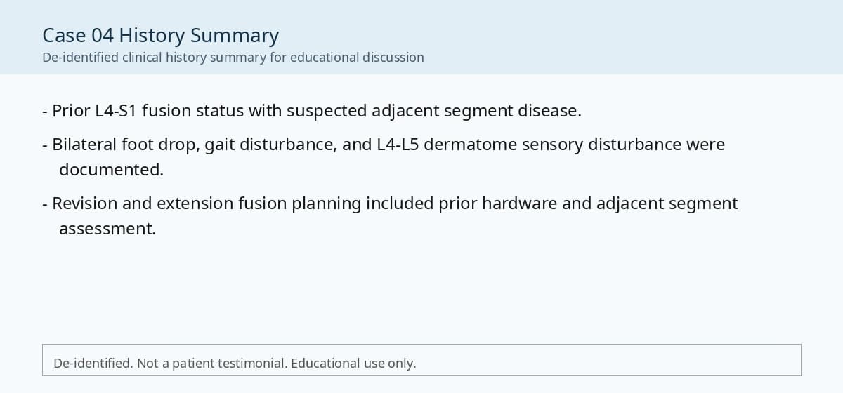
The patient presented with bilateral foot drop, gait disturbance, and sensory disturbance involving the L4-L5 dermatomes. The symptoms developed in the setting of previous lumbar fusion and were evaluated as a possible adjacent segment disease pattern.
Neurologic findings
Preoperative neurologic examination demonstrated bilateral ankle dorsiflexion weakness, approximately grade 3, and bilateral great toe dorsiflexion weakness, approximately grade 3. Sensory disturbance was documented in the bilateral L4-L5 dermatomes. Gait disturbance was clinically significant.
Prior nonoperative treatment
Not reported in the available educational summary.
Imaging and clinical concordance
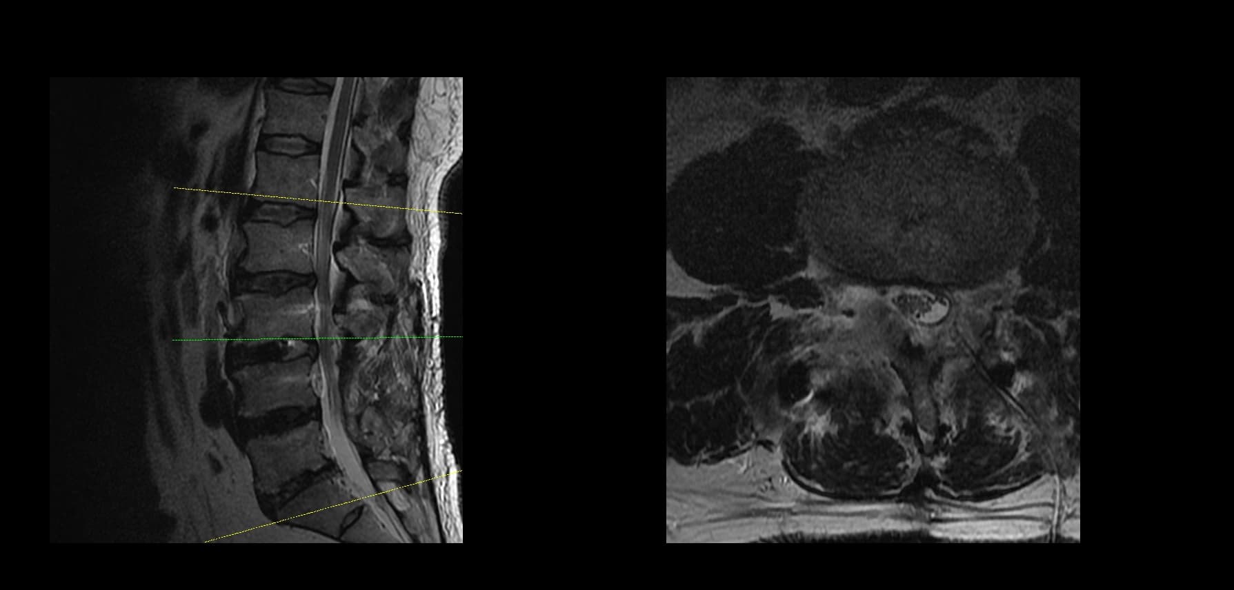
Preoperative lumbar radiographs and MRI demonstrated previous L4-L5-S1 fusion status with adjacent segment pathology at L3-L4. The L3-L4 level showed stenosis and neural compression concordant with the patient's bilateral neurologic symptoms. Dynamic radiographs and postoperative fusion status were reviewed as part of the revision and extension fusion planning.
Reasoning recorded for the procedure
The case was considered for revision and extension fusion because the patient had symptomatic adjacent segment disease above a prior fusion construct, with bilateral foot drop and imaging findings concordant with L3-L4 neural compression. The surgical objective was to decompress the affected neural structures and extend stabilization to the adjacent symptomatic level.
Operative considerations
UBE-assisted extension fusion was performed at L3-L4 in the setting of a previous L4-L5-S1 fusion construct. The procedure included careful exposure and dissection around the previous screw heads, screw removal or revision as required for extension of the construct, endoscopic decompression, disc space preparation, interbody fusion, cage placement, and posterior fixation. Particular attention was given to identifying the prior hardware safely and minimizing unnecessary tissue disruption during screw-head exposure and removal.
Postoperative course
Postoperative neurologic follow-up documented improvement of the bilateral motor deficit from approximately grade 3 to grade 5. This postoperative course is presented as an observation from this de-identified case only and should not be interpreted as predicting similar recovery in other patients.
Postoperative imaging
Postoperative radiographs demonstrated extension fusion involving the adjacent L3-L4 level in relation to the prior L4-L5-S1 fusion construct.
Educational point
This case illustrates surgical decision-making in adjacent segment disease after previous lumbar fusion. In revision and extension fusion, the operative target should be supported by symptoms, neurologic findings, prior fusion status, and imaging concordance. When fusion is performed in a spinal anesthesia environment, efficient operative workflow may be an important technical consideration. During screw removal or construct extension, careful dissection around the screw heads is essential to reduce unnecessary tissue trauma and to maintain a controlled revision corridor.
Published images and video
Media are identified by their public source. Journal figures open at the original source; operative video illustrates a technical example. Imaging and operative appearances do not measure pain relief, function, or durable fusion.
Clinical history
preoperative imaging
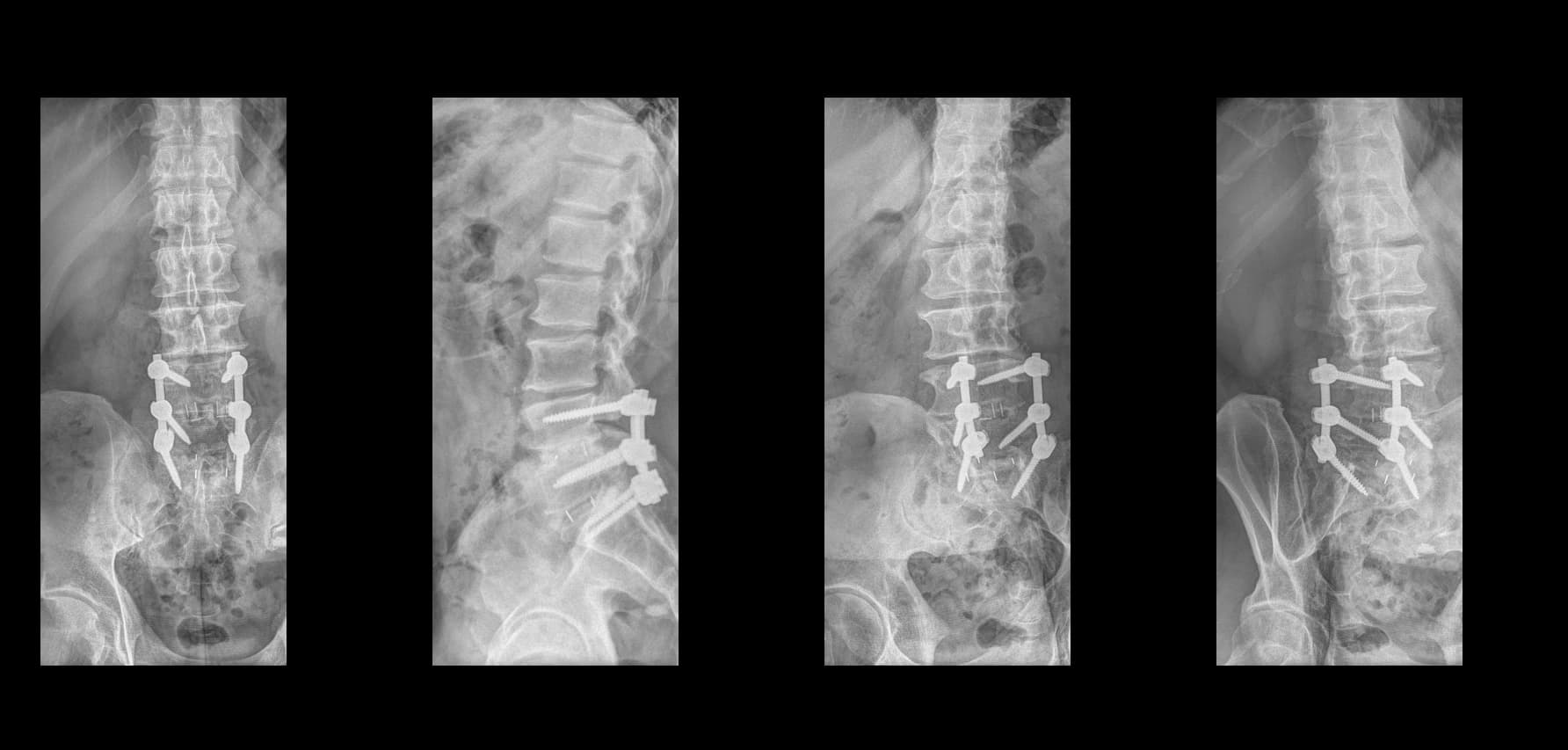
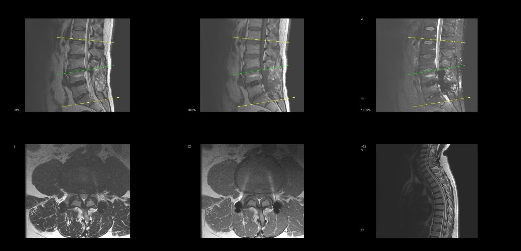
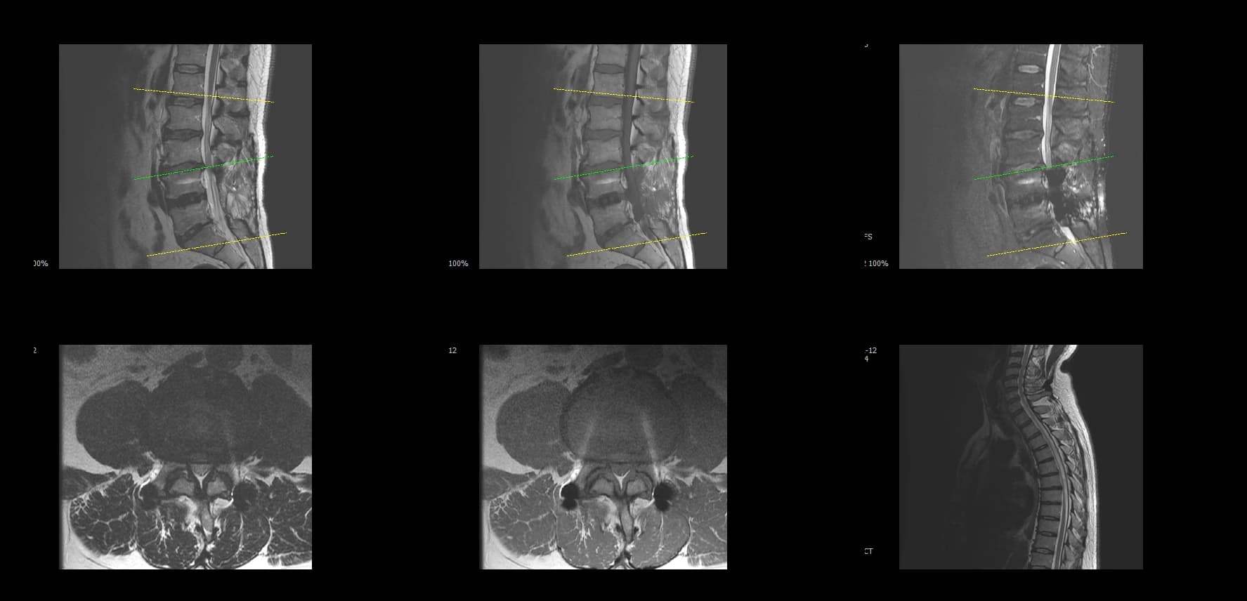
postoperative imaging
Reported observations and missing data
Values below are limited to what the identified public source reports; source charts were not re-audited for this website update. Operative-time definitions and observation windows may differ. Do not compare these values across surgeons or procedures.
- Operative time
- 65 minutes.
- Estimated blood loss
- Approximately 300 mL.
- Hospital stay
- 5 days.
- Complications
- No perioperative complication was documented in this educational case summary.
- Follow-up interval
- Not reported in the available educational summary.
- Validated patient-reported outcome scores
- Not reported in the available educational summary.
Evidence and further reading
These sources provide clinical context. They are separate from the source records for this case and do not verify its outcome.
Coauthored publication · technical report
Biportal Endoscopic Spinal Surgery for Recurrent Lumbar Disc HerniationsChoi DJ, Jung JT, Lee SJ, Kim YS, Jang HJ, Yoo B. Clin Orthop Surg. 2016;8(3):325–329.
A published technical discussion of biportal revision discectomy, scar dissection, and preservation of remaining stabilizing structures.
Applicability: This report concerns recurrent disc herniation. It does not establish the benefit of revision fusion or validate outcomes in the cases on this website.
External evidence · randomized trial
Decompression alone or with fusion for degenerative lumbar spondylolisthesis (Nordsten-DS): five year follow-upKgomotso EL et al. BMJ. 2024;386:e079771.
At five years, decompression alone was non-inferior to decompression with instrumented fusion in the trial population.
Applicability: Patient selection and exclusions matter. These findings do not establish that every slip needs fusion, nor compare UBE-TLIF with all alternative techniques.
Source, authorship, and limitations
Source material: Hanjin Jang, MD academic case collection. Case 04 in the previously published Case-Based Education collection by Hanjin Jang, MD. A separate case-level clinical review date and public consent record are not reported in the available source. This entry was organized on 2026-09-11; this date records a website update, not a new clinical review.
Selection is educational. No population denominator, consecutive recruitment process, comparative group, or complete follow-up dataset is supplied. A missing complication field does not mean that no complication occurred. Published observations do not predict another patient's outcome.
No new private patient files were added in this update. Corrections and future clinical additions follow the editorial policy.
Suggested citation: Hanjin Jang, MD academic case collection. UBE Extension Fusion for Adjacent Segment Disease. Casebank, Case 04. Updated 2026-09-11. https://www.hanjinjangspine1.com/casebank/adjacent-segment-disease-extension-fusion